Electrical Cardiometry™
Continuous, non-invasive hemodynamic monitoring
Continuous, non-invasive hemodynamic monitoring
Electrical Cardiometry™, or EC™, is a completely non-invasive method for the continuous determination of various hemodynamically important parameters.
The parameters describe the cardiovascular system (blood flow, contractility, vascular resistance), the thoracic fluid status and the blood oxygenation. Electrical Cardiometry™ gives a comprehensive picture of the hemodynamic circulatory system – in less time than it takes to measure blood pressure!
Electrical Cardiometry™ as implemented in ICON® are U.S. market-released for use in adults, children and neonates.
EC™ requires the application of four adhesive sensors to the surface of the skin.
EC™ has been validated against gold standard methods such as thermodilution.
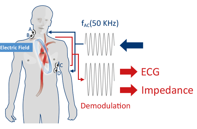
Placement of four skin sensors permits continuous measurement of changes of the thoracic electrical bioimpedance (TEB). By sending a low amplitude, high frequency electrical current through the thorax, the resistance (or impedance) which the current is facing (due to several factors) is measured. Application of advanced filtering techniques, Electrical Cardiometry™ (EC™) is isolating the impedance changes corresponding to varying blood flow.
The patented model of EC™ assumes that the underlying phenomenon of the characteristic change in thoracic impedance (or conductivity) during each cardiac cycle is the change in orientation of the erythrocytes (RBCs) when subjected to pulsatile flow.
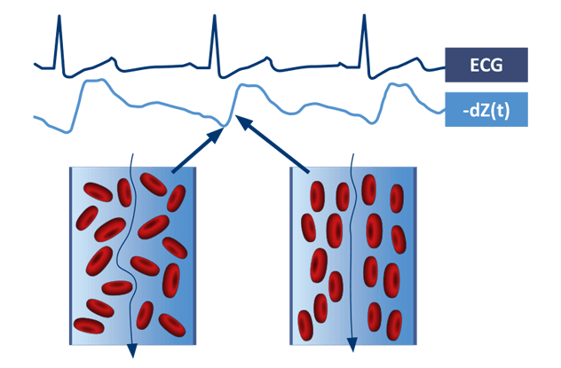
During diastole, the RBCs in the aorta assume a random orientation, which causes the electrical current to meet more resistance, resulting in a lower measure of conductivity. During systole, pulsatile flow causes the RBCs to align parallel to both the blood flow and electrical current, resulting in a higher conductivity state. By analyzing the rate of change in conductivity before and after aortic valve opening, or in other words, how fast the RBCs are aligning, EC™ technology derives the peak aortic acceleration of blood and the left ventricular ejection time (flow time). The velocity of the blood flow is derived from the peak aortic acceleration and used within Osypka Medical’s patented algorithm to derive stroke volume. Prior to measurement only weight, height, age and gender of the patient must be entered – that is all what is needed to start a measurement with an EC™ monitor.
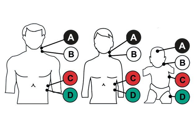
The EC™ Sensors must be placed at specific locations, as stroke volume measurement is depending on the volume of the electrically participating tissue, i.e., the thoracic volume.
The placement of the four sensors differs slightly depending on the age (or height) of the patient.
You need to load content from reCAPTCHA to submit the form. Please note that doing so will share data with third-party providers.
More InformationYou are currently viewing a placeholder content from Turnstile. To access the actual content, click the button below. Please note that doing so will share data with third-party providers.
More InformationYou are currently viewing a placeholder content from Facebook. To access the actual content, click the button below. Please note that doing so will share data with third-party providers.
More InformationYou are currently viewing a placeholder content from Instagram. To access the actual content, click the button below. Please note that doing so will share data with third-party providers.
More InformationYou are currently viewing a placeholder content from X. To access the actual content, click the button below. Please note that doing so will share data with third-party providers.
More Information
